More information on Hole-in-the-Head Disease
Main Ref: 41805  
Disease type: Parasitic infestations (protozoa, worms, etc.)  
Disease name: Hole-in-the-Head Disease  
Other names: Hexamita sp. Infection; Octomitus sp. Infection; Head and Lateral Line Erosion  
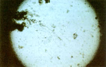
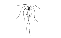
Caus. agent: Spironucleus sp.  
Group:    
Part of body and/or behavior affected by disease:
 Skins and/or fins  Belly  Swimming behavior
 Gills  Anus  Other behavior
 Eyes  Viscera Afflicted  Other symptoms
Fish affected:    
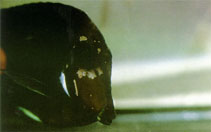
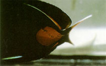
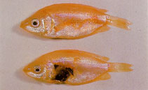
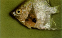
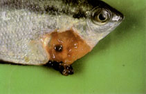
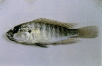
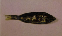
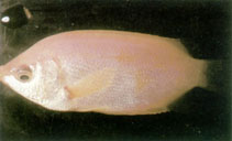
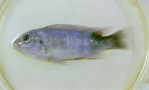
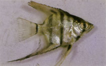
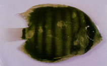
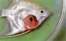
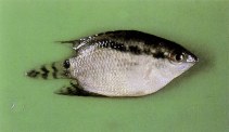
Symptoms: Symptoms include dark body color, emaciation, and white stringy excrements. In advanced infections, formation of reddish or pale patches on the skin and a secondary bacterial infection can occur. Excrements of the infected fish is a whitish-slime discharge that is cause by irritation or injury of the intestinal wall.  
Treatments: The use of Metronidazole for treatment has greater results than using Dimetridazole. Metronidazole Dose: 400-600 mg/100 liter for 3 days After this, aquarium water can be completely change or 50% waterchange combined with carbon filtration for at least 24-48 hours. In the food: mix 1 gm with 100 gm food and keep feeding this mixture for 3-5. Can also be mixed directly in the aquarium water when the fish has no appetite. Note: Effective for 3 days, but must be removed from the aquarium for it can cause damage to the kidneys and other internal organs. Control of secondary bacterial infections can be obtained by using anti-bacterial drugs such as Nitrofurazone (Dose: 10-15 mg/100 l for an unlimited time (or as instructed)) or Nifurpirinol (Dose: 250-500 mg/100 l for 3 days. At least 50% water change after treatment is recommended).  
Prophylaxis:    
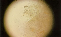
Remarks: These three flagellates that causes this disease are not easily distinguished, Spironucleus is common among tropical fish, Hexamita is associated with coldwaterfish and Octomitus has been found in Symphysodon discus. The 'holes' in the head are accumulation of problems (bad water conditions, unbalanced diet, and the parasites itself) which affect the lateral line organ . This results as a hole from which worm-like dots of rotten tissue protrude in the lateral lining in the head area.This hole symptoms can only be found in adult species. Also Ref. 41806 and 48502.  
Mortality and seasonality
Mortality:  
Seasonality:
Entered by: Palacio, Allan on 08.06.02
Modified by: Palacio, Allan on 01.07.04
Back to Search
tn_HoleHdD0.gif
picture (HoleHdD0.gif) by Bassleer, G.
tn_HoleHdD0.jpg
picture (HoleHdD0.jpg) by Bassleer, G.
tn_HoleHdD1.jpg
picture (HoleHdD1.jpg) by Bassleer, G.
tn_HoleHdD2.jpg
picture (HoleHdD2.jpg) by Bassleer, G.
tn_HoleHdD3.jpg
picture (HoleHdD3.jpg) by Bassleer, G.
tn_HoleHdD4.jpg
picture (HoleHdD4.jpg) by Bassleer, G.
tn_HoleHdD5.jpg
picture (HoleHdD5.jpg) by Bassleer, G.
tn_HoleHdD6.jpg
picture (HoleHdD6.jpg) by Bassleer, G.
tn_HoleHdD7.jpg
picture (HoleHdD7.jpg) by Bassleer, G.
tn_HoleHdD8.jpg
picture (HoleHdD8.jpg) by Bassleer, G.
tn_HoleHdD9.jpg
picture (HoleHdD9.jpg) by Bassleer, G.
tn_HoleHdDa.jpg
picture (HoleHdDa.jpg) by Bassleer, G.
tn_HoleHdDb.jpg
picture (HoleHdDb.jpg) by Bassleer, G.
tn_HoleHdDc.jpg
picture (HoleHdDc.jpg) by Bassleer, G.
tn_HoleHdDd.jpg
picture (HoleHdDd.jpg) by Bassleer, G.
tn_HoleHdDe.jpg
picture (HoleHdDe.jpg) by Bassleer, G.
tn_HoleHdDf.jpg
picture (HoleHdDf.jpg) by Bassleer, G.
tn_HoleHdDg.jpg
picture (HoleHdDg.jpg) by Bassleer, G.
tn_SpiInfD0.jpg
picture (SpiInfD0.jpg) by Bassleer, G.
Glossary
Comments & Corrections
     Sign our Guest Book

cfm script by eagbayani, 10.05.99 ,  php script by kbanasihan 05/27/2010 ,  last modified by sortiz, 06.27.17