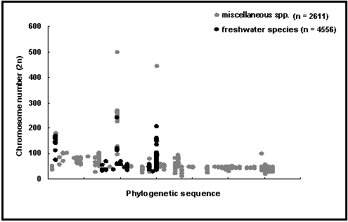
Karyological and cellular DNA content data (see Fig. 56) are important for studies of the genetics and systematics of fishes.
Locality: Refers to where the samples used were collected.
Country: Refers to the country of the sampling locality.
Sex: Refers to sex of samples used (unsexed, female, male or mixed).
Tissue(s) Used: Refers to tissue(s) used for the chromosomal study.
Chromosome number: Fields are provided for the haploid/gametic and the diploid/zygotic chromosome number. If the chromosome number is variable, the range is provided in the diploid/zygotic chromosome number fields.

Fig. 56. Chromosome number of freshwater fishes compared with that of miscellaneous species arranged in phylogenetic sequence from primitive (left) to modern (right). Note the decrease in chromosome decrease in chromosome number and variance for modern groups. See Box 34 for a discussion of this graph.
Chromosome types: Gives the numbers of chromosomes of different types:
metacentric: chromosomes whose centromeres are approximately midway between each end, thereby forming two chromosome arms of similar length;
submetacentric:
chromosomes whose centromeres are not at the middle of the chromosome (ratio of long arm to short arm is approximately 2:1);subtelocentric: chromosomes with a more terminally placed centromere, forming very unequal chromosome arms (ratio of long arm to short arm is approximately 3:1);
telocentric/acrocentric: chromosomes whose centromeres appear to be at the very tip of the chromosome;
meta-submetacentric: metacentric and submetacentric chromosomes.
subtelo-acrocentric: subtelocentric and acrocentric chromosomes.
Chromosome arm number: Gives the total number of chromosome arms, which is largely dependent on the chromosome types (e.g., a metacentric chromosome will have two arms while a telocentric chromosome will only have one).
Sex-determining mechanism: Gives information on how males and females of the species are designated (choices include xx-xy, xx-xo, etc. for those with sex chromosomes or no sex-associated heteromorphic chromosomes).
Genetic marker(s): States whether genetic marker(s) exist in the species and the choices are yes and no. A marker is a phenotypic characteristic (e.g., allozyme, chromosome band, etc.) that can be used to infer the genotype of an organism.
DNA content: Gives the specific haploid cellular content (in picograms). If references exist with values different from those in this field, they are placed in the remarks field.
Remarks: For miscellaneous comments, e.g., presence of structural rearrangements, specialized chromosomal features, sex-determining mechanism, polyploidization and, if any, other morphological markers.
To date, the GENETICS table covers more than 2,500 species with information extracted from over 2,200 references.
We used published references, checklists of chromosome numbers and karyotypes of different groups of fish aside from the database of Dr. Victor Arkhipchuk (1999) of Ukraine. Major sources include the Fish Chromosome Atlas of the National Bureau of Fish Genetic Resources (India) NBFGR (1998) and Klinkhardt et al. (1995).

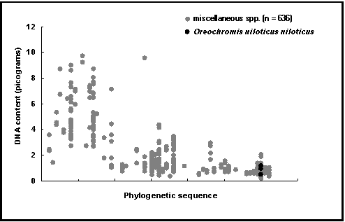
Fig. 57. DNA cell content of Oreochromis niloticus niloticus and miscellaneous species. Note that the decrease in DNA content from primitive (left) to modern groups (right) is similar to the independent decrease in chromosome numbers (Fig. 56).

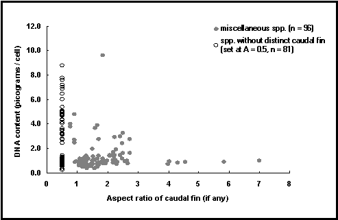
Fig. 58. DNA cell content as a measure of cell size vs. aspect ratio of caudal fin (A) as a measure of activity. See Box 34 for a discussion of this graph, and Fig. 52 for definition of the aspect ratio of caudal fins.
The DNA (deoxyribonucleic acid) content of plant and animal cells is extremely variable and few generalizations have emerged which can be used to predict the amount of DNA in the cells of a given group of organisms.
The most powerful of the existing generalizations is that the DNA content of cells tend to vary with cell size, suggesting a rough proportionality between the amount of DNA per cell, and the amount of living cellular material involved in various syntheses controlled by that DNA.
This generalization implies essentially that DNA content per cell, as recorded in the relevant field of the GENETICS table is a measure of cell size (see Cavalier-Smith 1991).
Given the tendency for organisms with large cells to have low metabolic rates, and conversely (von Bertalanffy 1951), animals with large cells (e.g., lungfishes, which reduce their metabolic rate during aestivation) will tend to have lots of DNA per cell (Thompson 1972).
In fishes, there is a clear pattern for chromosome numbers and for DNA (and hence cell size) to decline with derivedness, with perch-like fishes (high order number in Nelson’s (1994) classification) exhibiting a much lower range of DNA contents than more generalized, primitive forms (Hinegardner and Rosen 1972 and see Fig. 57). [Note that chromosome number and DNA content are not correlated, as indicated by Cavalier-Smith (1991) and confirmed by a FishBase graph not reproduced here.]
This may be thought to be the result of metabolic constraints, with fish cell size (and thus DNA content) declining with the evolution of high metabolic performance, such as displayed, e.g., by tunas (Cavalier-Smith 1991).
However, as also pointed out by Cavalier-Smith (1991), there is a lower limit to the size of cells: the fact that capillaries (which are formed by single cells) cannot have a diameter much smaller than that of red blood cells.
Combining all the above, one can hypothesize that a plot of DNA content vs. the caudal aspect ratio of fish (an index of metabolic intensity, see the SWIMMING table) should have on the left side of the plot a wide range of DNA content associated with low aspect ratios (including aspect ratio set at 0.5, to represent fish which do not use the caudal fin as their main organ of propulsion, and which tend to have low metabolic rates), and, on the right side of the plot, a narrow range of (low) DNA content associated with high aspect ratios. Fig. 58 displays these features, thus corroborating hypotheses linking DNA content¾
via cell size¾
to metabolic rate.
References
Cavalier-Smith, T. 1991. Coevolution of vertebrate genome, cell and nuclear sizes, p. 51-86. In G. Ghiara et al. (eds.) Symposium on the evolution of terrestrial vertebrates. Selected Symposia and Monographs. U.Z. I. 4, Modena.
Hinegardner, R. and D.E. Rosen. 1972. Cellular DNA content and the evolution of teleostean fishes. Am. Nat. 106(951):621-644.
Nelson, J.S. 1994. Fishes of the world. 3rd ed. John Wiley and Sons, Inc., New York. 600 p.
Thompson, K.S. 1972. An attempt to reconstruct evolutionary changes in the cellular DNA content of lungfish. J. Exp. Zool. 180:
von Bertalanffy, L. 1951. Theoretische Biologie Vol. II. A Francke A.G. Verlag, Bern. 418 p.
Daniel Pauly, Christine Casal and Maria Lourdes D. Palomares
362-372.
Clicking on the Biology button in the SPECIES window, then the Genetics button in the BIOLOGY and the following window will bring you to the GENETICS table.
On the Internet, you get to the GENETICS table by clicking on the Genetics link in the ‘More information’ section of the ‘Species Summary’ page. You can create a list of all species with available data by selecting the Genetics radio button in the ‘Information by Topic’ section of the ‘Search by FishBase’ page.
We thank P. Yershov and V. Arkhipchuk for their advice on the structure and content of this table.
Arkhipchuk, V.A. 1999. Chromosome database. (given to us in electronic file).
Klinkhardt, M, M. Tesche and H. Greven. 1995. Database of fish chromosomes. Westarp Wissenschaften. Germany. 237 p.
NBFGR. 1998. Fish Chromosome Atlas. National Bureau of Fish Genetic Resources Special Publication, No. 1. Lucknow, India. 332 p.